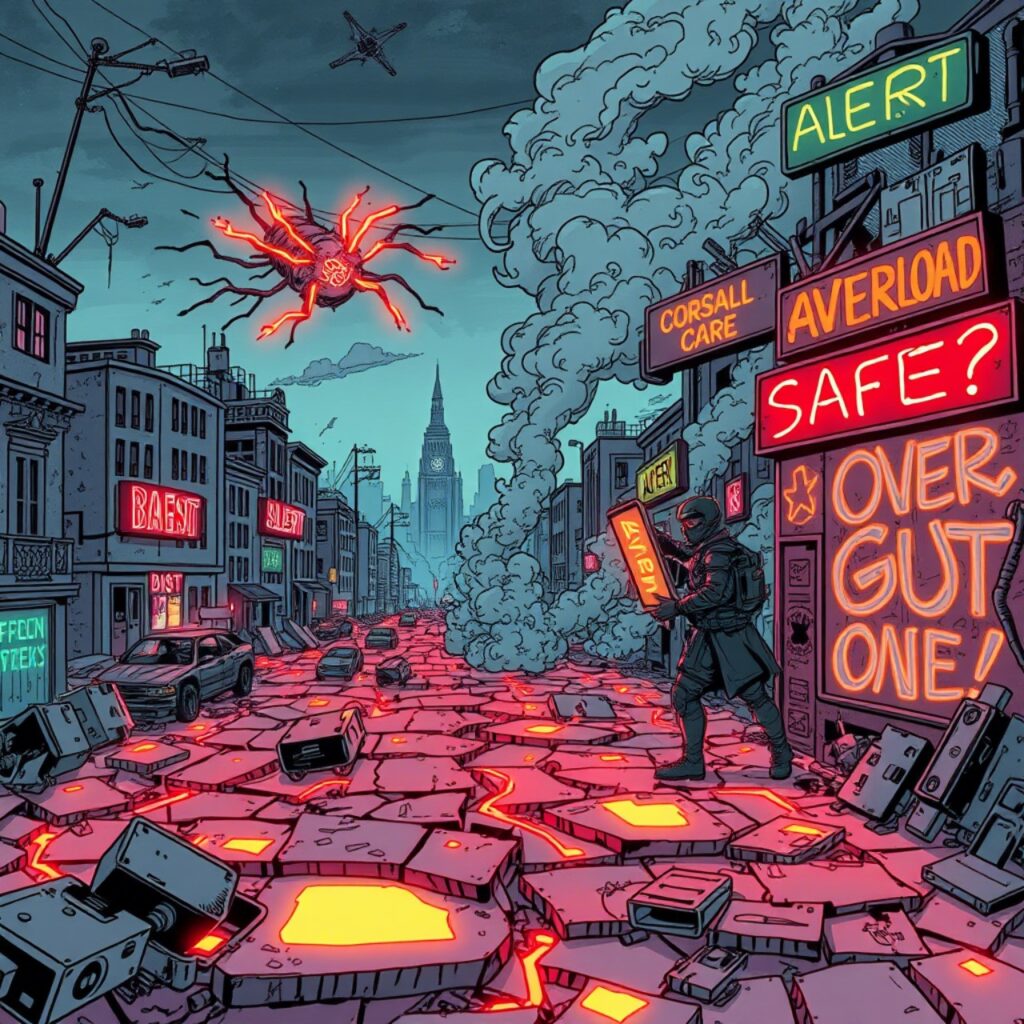
Microglia & Mast Cells: The Brain’s Fire Alarms and the Gut’s Riot Police

(Why Safety Can’t Return Until They Stand Down) 🔥 A System on High Alert
🔥 A System on High Alert
What do a hyperactive child, a shut-down autistic toddler, and a vaccine-injured adult with chronic fatigue have in common?
Their inner defense system never got the message that the war is over.
This page traces the cellular players responsible: microglia in the brain, and mast cells in the gut and body. These immune sentinels respond to infection, stress, toxins, and even perceived danger signals from cellular debris and ATP. In trauma, spectrum conditions, and chronic inflammation, they don’t switch off.
🧠 Microglia: The Brain’s Immune Sculptors
- Microglia make up ~10% of all brain cells.
- In early life, they prune synapses, guide myelination, and support neurodevelopment.
- When activated, they release inflammatory cytokines—TNF-α, IL-1β—and alter neurotransmitter levels.
In autism and ADHD:
- Microglia often show chronic activation even in the absence of infection.
- PET scans and postmortem studies reveal glial overgrowth, cytokine overload, and altered pruni
- ng patterns (Suzuki et al., 2013).
- Overactivated microglia disrupt myelination, leading to attention issues, poor auditory/language processing, and even social apathy.
“They’re supposed to garden the brain. Instead, they’re throwing Molotovs.”
💥 Mast Cells: The Gut’s Riot Squad
- These cells reside along mucosal surfaces—gut, lungs, skin, brain.
- They release histamine, serotonin, tryptase, prostaglandins—many of which affect mood, attention, and behavior.
- Mast cells talk constantly with neurons and microbes—and when the gut terrain is broken, they go rogue.
In ASD, ADHD, and PANS/PANDAS:
- Mast cell counts are elevated (Theoharides et al., 2016).
- Triggers include food antigens, toxins, stress, mold, and even non-infectious inflammation.
- Symptoms include mood instability, brain fog, hyperactivity, and physical symptoms like flushing or gut pain.
Many children are diagnosed with behavioral disorders when, in truth, they are living in a permanent immune thunderstorm.
- These cells reside along mucosal surfaces—gut, lungs, skin, brain.
🧬 The Vicious Cycle: Microglia ↔ Mast Cells ↔ Gut ↔ Brain
These two cell types talk to each other:
- A gut trigger (like LPS or spike protein) can activate mast cells ➝ releasing IL-6 and TNF-α.
- These cytokines cross into the brain (especially with a leaky BBB) and activate microglia.
- Microglia then further dysregulate dopamine, serotonin, and GABA, feeding the ADHD/autism cascade.
- The brain’s panic signal tells the gut it’s unsafe ➝ mast cells flare again.
This isn’t a glitch. It’s a feedback loop of survival mode—at the cost of developmental peace
📚 Evidence Base
- Suzuki et al., 2013 (JAMA Psychiatry): “Increased binding to TSPO receptors in the brains of children with ASD indicates microglial activation.”
- Theoharides et al., 2016 (J Neuroinflammation): “Mast cell activation contributes to neuropsychiatric symptoms via BBB disruption.”
- Estes & McAllister, 2015 (Ann Rev Neurosci): “Neuroimmune abnormalities observed in ASD include excessive cytokines, altered glial development, and immune dysregulation.”
- Critchfield et al., 2011: Children with ASD showed elevated TNF-α and IL-1β in cerebrospinal fluid.
- Patterson, 2009: Maternal immune activation during pregnancy activates fetal microglia, altering brain wiring long before birth.
🧘♀️ Why This Matters for Healing
You can’t force calm with behavioral therapy when the cells that sense the world are still screaming.
Any terrain reset must:
- Quieten microglial fire (e.g., butyrate, resveratrol, lion’s mane, omega-3s).
- Rebuild mucosal layers to prevent mast cell triggers.
- Reintroduce safety signals—like SCFAs, GABA, and oxytocin.
- Balance the microbial terrain, especially keystones that produce calming metabolites.
💡 Clinical Signals
- Families often notice sudden “breakthroughs” after anti-inflammatory interventions, mold detox, or microbiome resets.
- Children with ASD sometimes show a “spike window” after fever—possibly because immune cells shift phenotype during the febrile response.
In PANDAS/PANS, antibiotics or anti-inflammatories often restore function quickly, suggesting terrain-level immune chaos—not fixed brain dysfunction.
🔐 Closing Thesis
Until microglia and mast cells believe the war is over, the body won’t permit full connection.
This isn’t bad wiring. It’s a false alarm that no one turned off.
