Nedergaards’s glymphatics and neurodivergent expression
Nedergaards’s glymphatics and neurodivergent expression
Understanding how it explains the expression and the developmental stage collapse
How a disrupted gut ecosystem, fading acetylcholine, and lost deep sleep may help explain the modern wave of neuroinflammatory conditions
Most medical revolutions do not begin in a laboratory.
They begin with a question that refuses to leave you alone.
Mine began with a jar of beet kvass on the kitchen counter — the liquid fizzing quietly, as if it knew something I did not. I was a father trying to understand why so many children’s brains seemed inflamed, why sleep failed them, why sensory systems overloaded, and why the same patterns kept appearing under different diagnostic labels across the lifespan. The more I read, the more the fragments refused to stay separate.
Genetics. Dopamine. Pesticides. Trauma. Inflammation. Gut health. Sleep. Heavy metals. Neurotransmitters. Each offered a partial truth. None explained the overlapping rise of autism, ADHD, bipolar disorder, schizophrenia, Parkinson’s disease and Alzheimer’s disease in the same historical window.
Then the pattern clarified.
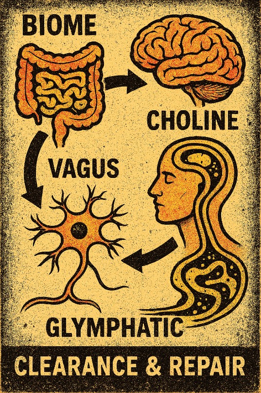
What if the final common pathway was not a single toxin or a single gene, but a failure of the brain’s nightly maintenance system? What if the glymphatic clearance machinery — the hydraulic network that moves cerebrospinal fluid through the brain during deep sleep — was being starved of the conditions it needs to operate? And what if that starvation began much further south, in the living ecosystem of the colon?
The idea was not mystical. It was mechanical. And once the pieces lined up, the timeline of modern neuroinflammatory conditions began to look less like a collection of unrelated epidemics and more like age-dependent expressions of a shared upstream collapse.
The cascade begins at the mucus layer
The story does not start in the neurons. It starts in a transparent gel lining the colon — the mucus bilayer that keeps the dense microbial community at a controlled distance from the epithelial cells beneath.
Modern industrial life has been hard on that layer. Antibiotics, ultra-processed foods, low fibre intake, disrupted early microbial exposure and chronic low-grade inflammation all chip away at the ecological engineers that help maintain it. Three bacterial groups have drawn particular attention: Akkermansia muciniphila, Faecalibacterium prausnitzii and members of the genus Roseburia. They are not the entire microbiome, nor are they universal heroes. Their value lies in the combination of roles they play in mucus interaction, fibre fermentation and short-chain fatty acid production, and in the frequency with which their abundance or activity appears altered in disease-associated profiles.
When this ecological niche is disturbed, the consequences ripple outward. Barrier signalling changes. Local immune tone shifts. The supply of microbial metabolites available to the host is altered. One nutrient pathway that sits at a particularly sensitive intersection is choline.
Choline is required for membrane phospholipids, for bile, and for the synthesis of acetylcholine — the neurotransmitter that supports parasympathetic signalling, including key aspects of vagal function. Experimental work has shown that selected gut bacteria can convert dietary choline into acetylcholine within the living intestinal environment, influencing local immune education. At the same time, inflammatory signals such as TNF-α and bacterial lipopolysaccharide can transcriptionally suppress the transporters that bring both free thiamine and thiamine pyrophosphate into colonocytes. Access, not merely dietary presence, becomes state-dependent.
The net effect is a plausible reduction in the chemical support for acetylcholine-dependent signalling. The vagus nerve — the major bidirectional highway between viscera and brain — begins to lose tone.
The switch that never fully flips
The vagus is often described as the “relaxation nerve.” That description is incomplete. It is one of the systems that helps shift the organism from high-alert sympathetic mode into the deep restorative physiology required for slow-wave sleep.
When cholinergic support weakens and inflammatory tone remains elevated, the transition into restorative sleep becomes less reliable. People still sleep. They still dream. But the architecture of that sleep can change. Slow-wave activity — the deep, slow, synchronised oscillations that coincide with the most robust glymphatic flow — becomes fragmented or reduced.
Research over the past several years has sharpened this picture. During non-REM sleep, coordinated oscillations in norepinephrine, cerebral blood volume and cerebrospinal fluid movement appear to act as a pump that drives fluid through the brain’s perivascular spaces. Astrocytic aquaporin-4 channels and vascular dynamics participate in the process. Human studies have begun to show that normal sleep, relative to sleep deprivation, is associated with higher morning plasma levels of Alzheimer-related biomarkers, consistent with overnight clearance into the periphery.
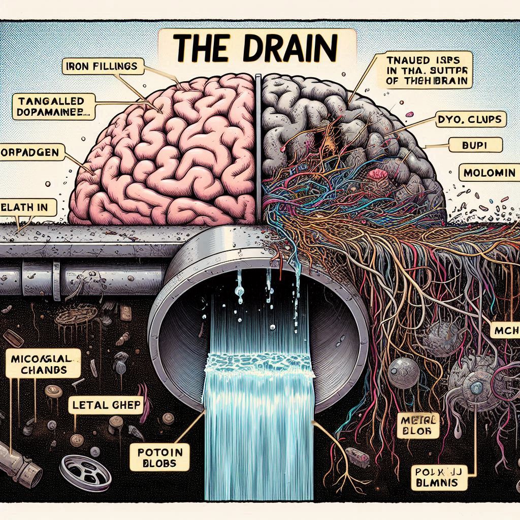
If the conditions required for that pump are repeatedly compromised, waste that the brain produces every day — oxidised metabolites, protein fragments, metals, inflammatory debris — is less efficiently removed. The internal environment of the brain slowly changes.
Waste that never fully leaves
The brain is a high-throughput organ. It generates metabolic by-products continuously. In a healthy system these are managed by a combination of cellular degradation, transport and fluid clearance. When nightly flow is impaired, the burden on the remaining systems rises.
Microglia — the brain’s resident immune cells — respond to the chemical environment around them. Persistent danger signals keep them in a pro-inflammatory state. Synaptic pruning becomes more aggressive. Sensory gain can increase. Networks that support exploration and flexible social cognition can be down-weighted in favour of protection and pattern detection. The brain is not “broken” in a simple sense. It is adapting to an internal environment that never fully resets.
The clinical expression of this adaptation depends heavily on developmental timing.
In the earliest years, when the brain is under rapid construction, a sustained shift in microglial and clearance dynamics can warp developmental trajectories — language, sensory processing, social reciprocity, sleep architecture. Later in childhood, when prefrontal circuits that support attention and working memory are under heavy demand, the same upstream constraints can present as difficulties with focus and regulation. In adolescence and early adulthood, when dopaminergic and limbic systems are both highly active and still maturing, the system can tip into mood instability or psychosis. In later life, the slow accumulation of uncleared material and the progressive weakening of the same circuits produce the more familiar neurodegenerative pictures.
Different ages. Different regional vulnerabilities. One recurring upstream signature: compromised conditions for nightly brain maintenance.
Clues that pointed the way
Several long-standing observations take on new coherence once clearance and cholinergic support are placed at the centre.
The strikingly high rates of nicotine use across schizophrenia, ADHD, bipolar disorder and, in some studies, autism spectrum conditions are one such clue. Nicotine is a nicotinic acetylcholine receptor agonist. For a brief window it can partially compensate for reduced cholinergic signalling — sharpening focus, damping sensory overload, transiently supporting vagal-related functions. It is not a cure. It is a chemical patch for a system that has lost part of its natural support.
The regional accumulation of metals in certain brain areas, repeatedly measured in both autistic children and older adults with neurodegenerative disease, is another. Metals are not necessarily the primary cause. They are also evidence of incomplete clearance. A system that cannot flush its interstitial spaces nightly will retain what a healthy system would remove.
Even the temporary improvements sometimes seen with agents that quiet extracellular ATP signalling (a danger signal to microglia) fit the pattern. When the immune cells are temporarily calmed, function can improve for weeks. When the underlying clearance deficit continues, the danger signals return and the window closes. The drug revealed the lock; it could not restore the upstream conditions that keep the lock from engaging.
The logic of repair
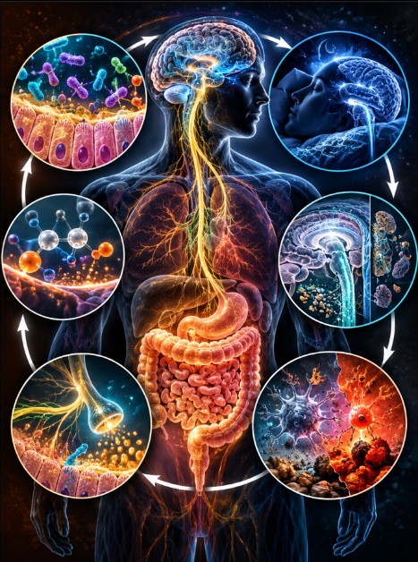
If the collapse follows a sequence — terrain disruption, altered choline and cholinergic support, reduced vagal and autonomic flexibility, fragmented deep sleep, impaired glymphatic dynamics, persistent microglial activation — then restoration is unlikely to succeed by attacking the final symptoms in isolation.
The logic points upstream.
Rebuilding microbial and mucus ecology. Supporting the metabolic pathways that feed acetylcholine and membrane integrity. Restoring conditions that allow the autonomic nervous system to shift more reliably into restorative states. Protecting and lengthening the windows of slow-wave sleep. Only then does the hydraulic machinery of brain clearance have a realistic chance of re-engaging. And only when the nightly backlog begins to ease do microglia receive the “all-clear” signals that allow them to shift toward repair rather than constant defence.
This sequence is not a treatment protocol. It is a research and clinical hypothesis about order of operations. Individual components already have varying levels of evidence behind them. The full causal chain, and the degree to which restoring it can reverse established developmental or neurodegenerative trajectories, remains to be tested rigorously.
A different kind of map
What has changed is the level of the question.
For decades the field has hunted for the gene, the toxin, the receptor, the protein that would explain each diagnostic category in isolation. The Secondary Biological State perspective, and the more specific TCVG relationships that sit inside it, suggest that some of the most important action may lie in the shared operating conditions of the system rather than in any single broken part.
The brain was never meant to function indefinitely without a nightly reset. When the conditions for that reset are chronically undermined, the organism adapts. The adaptations look different at age three, at age nine, at age twenty-two, and at age seventy. The underlying constraint — a system that cannot fully clear and restore itself — can be recognisably related.
This is not a claim that every case of every condition follows this exact path. Biology is more varied than that. It is a claim that a coherent, testable, systems-level mechanism now exists that can organise observations previously kept in separate silos.
The map is still incomplete. The measurements are still imperfect. The interventions still need careful evaluation. But for the first time the fragments are no longer forced to compete with one another. They can be read as chapters of the same story: terrain, acetylcholine support, autonomic flexibility, deep sleep, fluid clearance, and the immune cells that decide whether the internal environment is safe enough to rebuild.
When the brain stops cleaning itself at night, the world records the consequences as disease.
When the conditions for cleaning are restored, the same biology that produced the symptoms begins, slowly and imperfectly, to remember how to reverse them.
It is not magic.
It is hydraulics, chemistry, ecology and sleep — working, or failing to work, in sequence.
That is the hypothesis.
Now it needs to be tested with the seriousness it deserves.