
THE SIGNAL AND THE SILENCE

The Signal and the Silence was written during the first phase of our investigation into the relationship between the microbiome, immune regulation and neurological function. It contains hypotheses and interpretations that have since evolved, but it also records the questions and connections that drove the work forward.
This article is a condensed version of that investigation. The original book, including its full diagrams, references and extended arguments, is available here.
THE SIGNAL AND THE SILENCE
How the collapse of three gut microbes opened a window onto the immune roots of modern neurological and inflammatory disease
There is a pattern that keeps appearing in the research, quiet at first and then impossible to ignore.
Across dozens of studies, children on the autism spectrum, adults with inflammatory bowel disease, patients with multiple sclerosis, long COVID, and chronic fatigue show the same microbial signature: a marked reduction or near-absence of three keystone species — Akkermansia muciniphila, Faecalibacterium prausnitzii, and Roseburia. These are not obscure organisms. In a healthy colon they are among the most abundant and functionally important residents. Their consistent disappearance across conditions that medicine still treats as separate raises a question that is both simple and unsettling.
What happens when the microbes that hold the intestinal wall together, produce the metabolites that calm inflammation, and help train the developing immune system are no longer there?
The answer is not a single disease. It is a cascade.
The Three That Held the Line
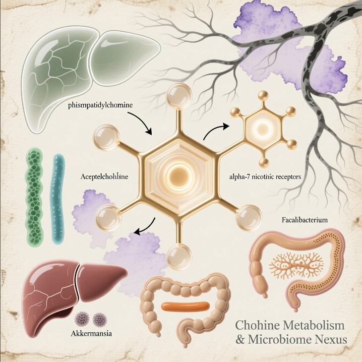
Akkermansia muciniphila lives in the mucus layer that lines the gut. It consumes host mucin and, in doing so, stimulates the production of more mucus, maintains barrier integrity, and releases outer-membrane proteins that engage the immune system toward tolerance rather than alarm. Faecalibacterium prausnitzii is one of the most potent anti-inflammatory organisms known in the human gut. It produces butyrate, the short-chain fatty acid that fuels colonocytes, supports regulatory T cells, and suppresses NF-κB-driven cytokine release. Roseburia species perform similar fermentative work, generating butyrate and propionate while helping regulate motility and energy signalling.
Together they form an ecological unit. Akkermansia produces acetate that cross-feeds the butyrate producers. Butyrate, in turn, helps maintain the low-oxygen environment and epithelial health that Akkermansia needs. When one declines, the others become more vulnerable. When all three fall, the colon loses its primary source of barrier reinforcement, local anti-inflammatory signalling, and metabolic support for the cells that line it.
This is not theoretical. Biopsy and sequencing studies have documented the loss repeatedly. In autism spectrum cohorts the depletion is often severe and appears early. The same pattern surfaces in ulcerative colitis, Crohn’s disease, Parkinson’s, type 2 diabetes, and post-viral syndromes. The organisms are not the only ones that change, but they are among the most consistent markers of a deeper ecological failure.
How the Line Breaks
The modern collapse of these species does not require exotic explanations. It follows the ordinary interventions of industrial life.
Cesarean delivery bypasses the maternal vaginal and fecal microbiota that normally seed the infant gut. Formula feeding removes the human milk oligosaccharides that selectively nourish early colonisers which later create conditions favourable to the butyrate producers. Broad-spectrum antibiotics, especially when given repeatedly in the first years of life, can reduce Faecalibacterium by more than 90 percent in experimental and clinical settings; recovery is often incomplete. Ultra-processed diets supply far less of the complex fibres these organisms require. Emulsifiers thin the mucus layer on which Akkermansia depends. Glyphosate, designed as an antimicrobial, shows particular activity against many anaerobic Firmicutes.
Each factor alone may be survivable. Together, and especially when they strike before the immune system has finished learning tolerance, they create a predictable outcome: a gut that is thinner, more permeable, less able to produce the short-chain fatty acids that keep local inflammation in check, and more likely to leak microbial fragments into systemic circulation.
The Immune System Loses Its Teachers
The infant immune system is not born knowing self from non-self. It is educated. Regulatory T cells, the peacekeepers that dampen unnecessary responses to food antigens and commensal microbes, are strongly induced by butyrate acting through receptors such as GPR109A. Dendritic cells are shaped by the same metabolites. The mucus layer itself keeps bacterial products from constantly triggering innate sensors.
When the trio is absent, that education is disrupted. Tight-junction proteins are less well maintained. Mucus production falters. Lipopolysaccharide and other microbial molecules reach the bloodstream more readily. The innate system registers danger. Without adequate butyrate, regulatory T-cell numbers and function decline while Th17 pathways become more dominant. The result is a state of low-grade, directionless vigilance — chronic activation without clear resolution.
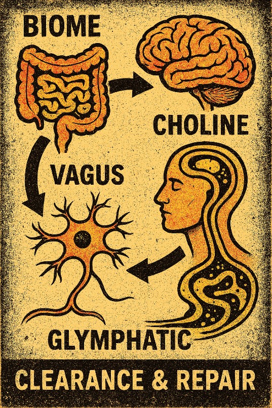
This inflammatory tone does not stay in the gut. Cytokines travel. Microglia, the brain’s resident immune cells, respond to the same signals. In a developing brain they can alter synaptic pruning, shift excitatory-inhibitory balance, and change the way sensory information is filtered. The same axis that links gut barrier failure to systemic inflammation also links it to changes in neurotransmitter systems: tryptophan is diverted toward the kynurenine pathway under inflammatory pressure; dopamine and GABA signalling are modulated by short-chain fatty acids and by the integrity of the blood-brain barrier; cholinergic tone, already sensitive to choline availability and membrane status, is further constrained by inflammation at nicotinic receptors.
The book that first mapped this territory framed the outcome starkly: autism as the neurological signature of an early immune misfire driven by microbial collapse. The language is strong. The underlying observations are more measured. Correlations between early microbiome disruption and later neurodevelopmental outcomes are now extensive. Interventional signals — temporary symptom reduction after Suramin in a small open-label trial, sustained improvements in some children after intensive fecal microbiota transplant protocols — exist and deserve rigorous follow-up. Causation, however, remains unproven. Autism is multifactorial; genetics, prenatal environment, and other factors still matter. What the microbial pattern supplies is not a complete explanation but a coherent, testable entry point into the immune and metabolic dimensions of the condition.
Metabolic and Neurotransmitter Fallout
The same organisms that regulate immunity also shape energy metabolism. Butyrate activates pathways of mitochondrial biogenesis. Acetate contributes to ketone production. When these metabolites decline, colonocytes and other cells shift toward less efficient glucose oxidation, reactive oxygen species rise, and metabolic flexibility is lost. Choline metabolism is altered as well. In a dysbiotic environment, choline can be diverted by certain bacteria toward trimethylamine and then to TMAO, while host tissues experience relative shortfall in a nutrient required for acetylcholine, phosphatidylcholine membranes, and one-carbon metabolism.
The neurotransmitter consequences follow. Serotonin synthesis is pulled toward neurotoxic kynurenine metabolites under inflammatory pressure. Dopamine signalling is blunted by reduced receptor expression and barrier leak. GABA tone falls when the microbes that support inhibitory signalling are missing. Vagal afferent signalling, which depends in part on short-chain fatty acids and intact mucosal architecture, weakens. The brake pedal on stress and inflammation becomes less effective.
None of these changes requires the microbes to manufacture human neurotransmitters directly. They change the chemical and structural conditions under which host signalling operates. That is often enough.
Sensory Gating and the Adaptive State
Sensory gating — the brain’s ability to filter irrelevant stimuli — depends on GABAergic inhibition, cholinergic modulation, and intact thalamic circuitry. When inflammatory cytokines rise and short-chain fatty acids fall, filtering weakens. The result is a brain that receives more raw data than it can organise. Lights are brighter, sounds sharper, textures more intense. Gaze aversion, stimming, and withdrawal can be understood, in this light, as adaptive responses to signal overload rather than primary deficits of motivation or social interest.
This framing does not romanticise autism or deny its challenges. It simply relocates part of the mechanism from an exclusively genetic or purely psychiatric domain into a neuroimmune and ecological one. If the underlying drivers are inflammatory and metabolic, then interventions that restore barrier function, short-chain fatty acid production, and regulatory immune tone become rational targets for research — not guaranteed cures, but plausible levers.
The Return Path
Restoration is not achieved by swallowing a capsule of the missing species into a hostile environment. The terrain must first become permissive again. That means reducing ongoing sources of mucosal injury — emulsifiers, residual antibiotics, excessive oxidative load — while supplying the substrates the keystone organisms require: specific polyphenols that favour Akkermansia, resistant starches and diverse fibres that feed the butyrate producers, and conditions that allow oxygen gradients and mucus production to normalise.
Clinical signals already exist. Structured fecal microbiota transplant protocols have produced measurable shifts in the target taxa and correlated behavioural improvements that persisted for years in some cohorts. Smaller nutritional and prebiotic interventions show more modest but consistent movement in the same direction. The timeline is measured in months, not days, because epithelial renewal, immune recalibration, and ecological reassembly are slow processes.
What the Pattern Actually Proves
The strongest claim the data currently support is not that the loss of three microbes “causes” autism or any other single diagnosis. It is that their depletion is a shared, early, and functionally consequential feature of multiple modern chronic conditions. The same organisms that maintain the gut barrier, generate the metabolites that restrain inflammation, and help calibrate the developing immune system are repeatedly absent in diseases characterised by barrier failure, chronic immune activation, and altered neural signalling.
That pattern is robust enough to demand attention. It is precise enough to guide mechanistic research. And it is hopeful enough to justify careful, terrain-focused clinical exploration.
The microbes were never the whole story. They were the sentinels. When the sentinels fall, the signal changes. The silence that follows is not empty. It is the sound of a system operating under new, harsher constraints — constraints that, in principle, can be measured, understood, and, in time, relieved.