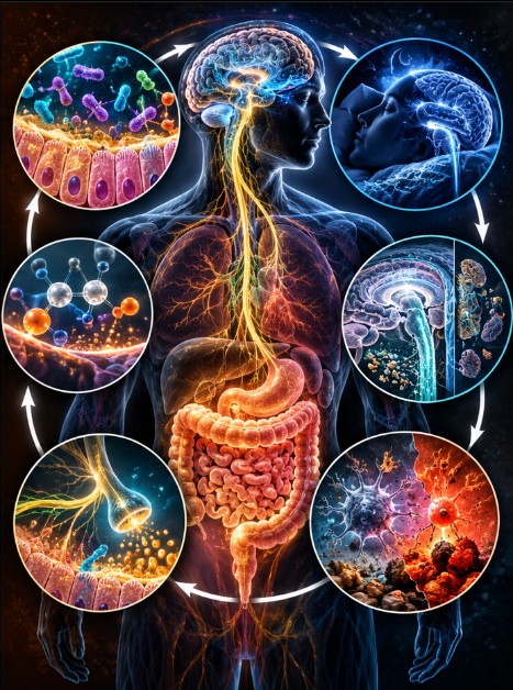
TCVG: The Biological Loop Connecting the Gut to the Brain
TCVG: The Biological Loop Connecting the Gut to the Brain
From the three gatekeepers of colonic barrier health to choline, vagal regulation and the brain’s maintenance system
An emerging systems hypothesis proposes that chronic biological dysfunction may be better understood not as a series of isolated failures, but as a disturbance in the feedback loops that connect the gut ecosystem, cellular metabolism, autonomic regulation and the brain’s capacity to maintain itself. After a year of intensive research and stress-testing, the TCVG framework offers a coherent architecture for these interactions—one that treats the body as a living, coupled system rather than a collection of separate parts.
The problem with looking at the body in pieces
Modern medicine has become extraordinarily good at examining individual components of human biology. We can measure inflammatory markers, sequence microbial DNA, image the brain, analyse metabolites and identify changes in neural activity. We can investigate the immune system, the autonomic nervous system, cellular energy production and the mechanisms through which the brain removes metabolic waste.
But a more difficult question remains: what happens when several of these systems begin to influence one another in a persistent cycle of dysfunction?
A disturbed gut ecosystem can change the metabolites available to the host. Host physiology, in turn, determines which microorganisms can survive and what they can do. Metabolic pathways influence membrane composition and neural signalling. Neural and autonomic activity regulate gut function. Sleep, circadian rhythms, vascular physiology and immune signalling influence the conditions under which the brain maintains its internal environment.
These are not separate conversations. They are overlapping parts of a living system.
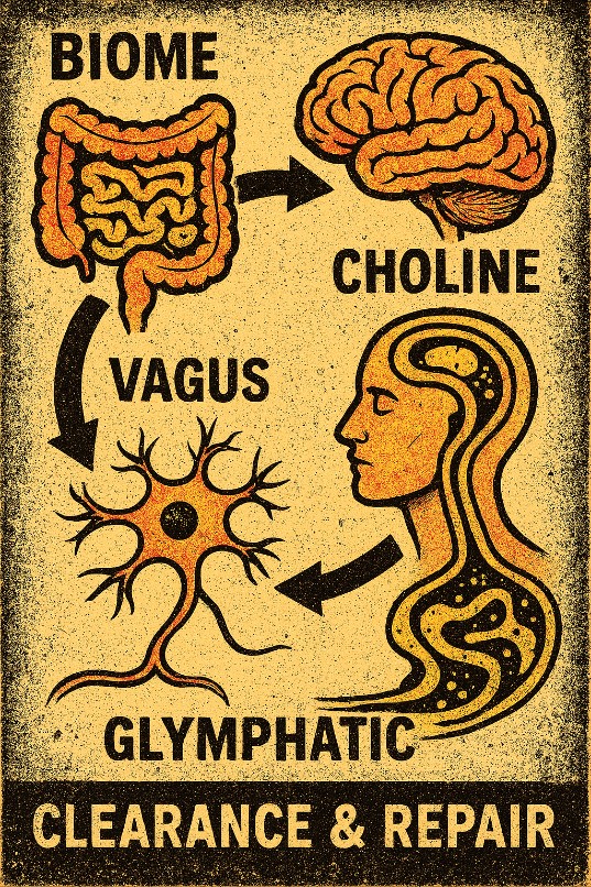
The TCVG model emerged from exploring these connections as a single biological architecture. Its four components are:
- T — The Trio and the wider gut biome
- C — Choline-dependent metabolic and signalling pathways
- V — The vagus nerve and autonomic regulation
- G — The glymphatic system and the wider machinery of brain maintenance
The model initially appears to describe a sequence: gut ecosystem, choline, vagal regulation, brain clearance. But that apparent linearity is misleading. Each component can influence the others, directly or indirectly, and the final component connects back to the conditions that shape the first.
TCVG is therefore best understood as a proposed coupled feedback system, not a one-way chain of causation.
The distinction matters. A linear model asks where a problem starts. A feedback model also asks why it persists, what reinforces it and what conditions might allow the system to recover.
T — The Trio: three gatekeepers of colonic barrier health
The first component begins in the colon, where the relationship between microorganisms and the host is particularly intimate.
The intestinal microbiome is a complex ecosystem containing bacteria, archaea, viruses, fungi and other biological components. These organisms perform overlapping functions involving the processing of dietary substrates, the production and transformation of metabolites, interactions with the immune system and the maintenance of the intestinal environment.
Within that much larger ecosystem, our framework focuses on three bacterial groups: Akkermansia, Faecalibacterium and Roseburia. We call them the Trio.
They are not the entire microbiome, nor are they responsible for every essential microbial function. Their significance lies in the particular roles they perform and in their potential value as indicators of changes in the wider ecosystem.
The intestinal barrier is not simply a wall separating the body from its contents. The colonic epithelium is a living layer, generally one cell thick, supported by mucus, tight junctions, immune defences and an intricate network of local signalling. Its job is not to exclude everything indiscriminately. It must allow useful substances to be absorbed while regulating contact with microorganisms, microbial products and other material in the intestinal lumen.
The Trio participates in different aspects of this relationship.
Akkermansia muciniphila occupies a specialised ecological niche involving mucin, an important component of the mucus layer. Its activity can contribute to microbial cross-feeding, in which the products generated by one organism become substrates for others. Its effects depend on the surrounding ecosystem: mucus degradation is not inherently beneficial or harmful in every context. Recent work continues to highlight its role as a key modulator of host physiology—strengthening barrier integrity, influencing metabolic health, and even showing promise in supporting weight-loss maintenance in clinical settings—while underscoring that context and microbial neighbourhood matter.
Faecalibacterium, including F. prausnitzii, and members of the genus Roseburia are important butyrate-producing bacteria. Butyrate is a short-chain fatty acid used extensively by colonocytes, the epithelial cells lining the colon. It also influences epithelial barrier biology, local immune signalling and the metabolic environment of the gut. These bacteria do not act alone; numerous other organisms contribute to butyrate production and the wider network of microbial metabolism. Emerging findings add further intrigue: certain Roseburia species, such as R. inulinivorans, have been linked to muscle strength and metabolic adaptations in both human associations and mouse models, suggesting the reach of these organisms extends beyond the gut wall itself.
Their functions make the Trio scientifically interesting. But the deeper hypothesis is about their ecological position.
First to go, last to return?
Across our developing review of microbiome research, we have repeatedly encountered reports of depletion or altered abundance involving members of the Trio in disease-associated microbial profiles. This has led us to a working hypothesis: some of these organisms may be among the early casualties of a disrupted intestinal ecosystem and among the slower populations to re-establish during recovery.
That is a hypothesis to test, not a universal rule. Microbial findings vary by disease, population, diet, medication, sampling method and study design. Relative abundance also does not necessarily tell us whether a microorganism is metabolically active or whether its function has been lost.
Nevertheless, the potential significance is considerable.
If the Trio is repeatedly altered across different conditions, it may offer a useful sentinel signal of ecosystem disturbance. It may tell us something about the environment in which the wider microbial community is attempting to function, even though no three organisms can fully represent that community.
In our own synthesis, we have developed provisional estimates suggesting frequent Trio alterations across the neurodevelopmental and autoimmune literature we examined. Those estimates remain internal working figures rather than independently verified pooled prevalence statistics. They require a transparent evidence table, defined inclusion criteria and formal replication before they can be treated as population-level findings.
The central question is not whether every person with a chronic condition lacks these three bacteria. It is whether recurring changes in their abundance or function reveal a broader pattern of disturbed host–microbe relationships.
The barrier is an ecosystem achievement
Barrier health is not supplied by a single bacterium. It emerges from interactions among mucus production, epithelial metabolism, tight junctions, immune tolerance, diet, microbial competition and the metabolites produced throughout the community.
When this relationship is disturbed, several changes may occur together: butyrate production may fall or change, epithelial energy supply may be affected, microbial metabolites may shift, and immune signalling may become less well regulated.
These effects are neither automatic nor identical in every person. Reduced abundance of a butyrate-producing organism does not prove that total butyrate production is low, just as a change in one metabolite does not establish that the entire barrier has failed.
The important point is that the host and its microbiome maintain a shared environment. Each influences the conditions required by the other.
The Trio provides our entry point into that relationship. TCVG asks what happens when disturbances in this ecological partnership extend beyond the colon.
C — Choline: the metabolic connection
The second component is choline, an essential nutrient involved in several distinct biological pathways.
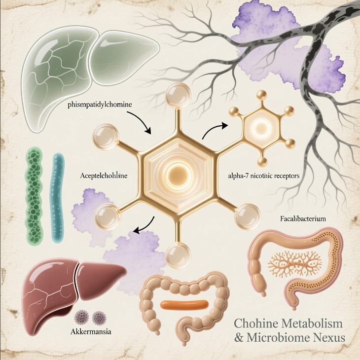
Choline contributes to the production of phosphatidylcholine, a major component of cellular membranes and an important constituent of bile. It can also be converted into acetylcholine, a neurotransmitter involved in neural communication, including autonomic signalling. Through its conversion to betaine, choline also participates in one-carbon metabolism and methyl-group transfer.
These functions make choline a plausible connection between intestinal physiology, cellular maintenance and nervous-system regulation.
But choline is not a single-purpose fuel, and its different pathways should not be collapsed into one simplistic mechanism.
Phosphatidylcholine contributes to membrane structure and lipid transport. Bile supports the digestion and absorption of fats, while bile acids also act as signalling molecules and undergo extensive microbial transformation in the intestine. Acetylcholine supports communication between neurons and is involved in parasympathetic and other cholinergic functions. Betaine contributes to methylation chemistry.
The body must maintain these activities within a changing physiological environment.
Where the microbiome enters the picture
The gut microbiome influences the availability and transformation of many dietary compounds. Some microorganisms consume choline; others produce metabolites that influence host metabolism. Microbial activity also affects bile-acid composition, intestinal inflammation and the environment in which nutrients are absorbed and processed.
Meanwhile, the host determines how much choline is supplied through diet, how it is absorbed and metabolised, how bile is produced and released, and how much demand exists for membrane synthesis and repair.
This creates several possible points of interaction between the Trio and choline-dependent physiology.
A disrupted intestinal ecosystem could alter microbial metabolism, bile-acid signalling and the inflammatory environment. Changes in host metabolism could, in turn, affect membrane maintenance, nutrient handling and the pathways supporting acetylcholine production.
Exciting recent work adds depth here. Stable-isotope studies have shown that the gut microbiome can contribute to de-novo choline synthesis from dietary substrates such as inulin, with labelled choline appearing in host organs including the brain. Other research has revealed that certain commensals can convert dietary choline into acetylcholine itself, influencing mucosal immune education, IgA responses and resistance to infection. These findings illustrate how microbial chemistry can feed directly into host cholinergic and immune pathways—precisely the kind of metabolic junction TCVG highlights.
What has not yet been established is that depletion of the Trio necessarily causes choline deficiency, or that the body automatically diverts choline away from acetylcholine whenever intestinal inflammation increases. Those are specific mechanistic propositions that require direct evidence.
The stronger starting point is that choline-dependent pathways sit at the intersection of membrane biology, metabolism, bile and neural signalling. Their status may therefore help us understand how a disturbance in one part of the system affects others.
In TCVG, choline is a potential metabolic junction—not a single switch that explains the entire system.
V — The vagus: a two-way communication network
The vagus nerve is one of the major communication routes between the internal organs and the brain. It carries sensory information from the body towards the central nervous system and contributes to the regulation of organs through autonomic output.
It is not the only route connecting the gut and brain. Spinal sensory pathways, endocrine signals, immune mediators, circulating metabolites and other neural circuits also participate. Nevertheless, the vagus is an important part of the communication architecture that makes gut–brain interaction possible.
Its relevance to TCVG becomes clearer when we stop thinking of the gut as a passive organ that sends occasional messages to the brain.
The intestine is continually reporting information about its internal conditions. Nutrient availability, distension, chemical signals, immune activity and microbial metabolites can all influence the information reaching the nervous system. The brain and autonomic nervous system then influence intestinal motility, secretion and other aspects of digestive physiology.
The conversation runs in both directions.
Acetylcholine and inflammatory regulation
Acetylcholine is a major neurotransmitter in parasympathetic signalling and is also involved in pathways through which neural activity can influence immune responses.
Experimental research into the cholinergic anti-inflammatory pathway has investigated how neural signalling, including signalling involving nicotinic acetylcholine receptors such as α7-nAChR, can modulate inflammatory activity. This is an important area of research, but the pathway is not a universal inflammation off-switch. Its operation depends on the cell types, tissues, receptors and conditions involved.
Within TCVG, this gives choline-dependent physiology and vagal regulation a plausible point of connection.
If metabolic conditions alter cholinergic signalling, or if autonomic regulation changes the intestinal environment, the effects could extend beyond a single organ. Changes in motility, secretion, barrier conditions and immune activity could alter the habitat experienced by the microbiome. Conversely, changes in microbial metabolism and gut signalling could influence neural and autonomic responses.
Cutting-edge evidence strengthens this picture. Direct recordings show that the gut microbiome regulates vagal afferent activity through metabolites such as short-chain fatty acids, bile acids and others; germ-free animals display reduced vagal tone that is restored by microbial colonisation. Nutrient sensing via the vagus can drive acetylcholine release in hippocampal circuits that support memory formation. Microbial ammonia has even been shown to enhance colonic acetylcholine levels and influence motility, revealing compensatory microbial mechanisms that respond to host cholinergic state. Meanwhile, resilient individuals appear to display both healthier gut-barrier microbial activity and more adaptive brain-network patterns linked to emotional regulation.
These interactions provide a biologically credible basis for studying the C–V connection.
They do not mean that every person with altered gut bacteria has vagal dysfunction, or that a measurement such as heart-rate variability can establish vagal failure on its own. Heart-rate variability is influenced by breathing, activity, medication, cardiovascular conditions and other factors.
The research question is more precise: when gut ecology and choline-dependent pathways change, do measurable alterations in autonomic regulation occur, and do those alterations help explain changes elsewhere in the system?
That is testable. It requires measurements across time rather than assumptions based on one symptom or one biomarker.
G — The glymphatic system: the brain’s maintenance architecture
This is where TCVG must go beyond the familiar idea of a gut–brain connection.
The glymphatic system is often introduced as a mechanism for clearing waste from the brain, particularly during sleep. That description is useful, but incomplete if it leaves the impression that the system is simply a drainage pipe attached to the end of a biological chain.
The brain is a metabolically demanding organ. Neurons continually generate electrical activity, maintain ion gradients, synthesise and recycle proteins, remodel synapses and communicate with glial cells. These processes produce metabolic by-products and require the extracellular environment to remain within tightly regulated limits.
The brain must manage water and solute movement, maintain ionic conditions, process cellular debris and remove or transform substances that accumulate during normal activity. It does so through several overlapping systems, including enzymatic degradation, cellular uptake, transport across the blood–brain barrier, microglial activity and fluid-associated clearance.
The glymphatic system is one component of this wider maintenance network.
More than waste removal
Research associated with Maiken Nedergaard and colleagues has helped establish the glymphatic framework: cerebrospinal fluid moves along pathways associated with cerebral blood vessels and exchanges with fluid in the brain’s interstitial spaces. Astrocytes, including the organisation of aquaporin-4 water channels at their vascular endfeet, are important to current models of this process.
The movement of fluid and solutes is influenced by several factors, including vascular pulsations, respiration, neural activity and sleep physiology. Recent work has sharpened the picture further: norepinephrine oscillations during non-REM sleep appear to drive rhythmic vascular dynamics that pump cerebrospinal fluid through the system. Human imaging studies are beginning to capture parallel signatures of large-scale CSF pulsations and inverse blood–CSF volume coupling during sleep. Evidence is also accumulating that sleep-active glymphatic processes can elevate morning plasma levels of Alzheimer-related biomarkers relative to sleep deprivation, consistent with overnight clearance to the periphery.
The details of glymphatic transport, its relative contribution to different clearance processes and the translation of findings from animal models to humans remain active areas of scientific investigation. Debates continue about the precise timing and relative importance of sleep versus wake states, yet the broader significance of coordinated fluid dynamics for brain homeostasis is increasingly clear.
The significance of this work extends beyond the removal of individual waste molecules.
The brain’s maintenance economy depends on a balance between what neural tissue produces, what cells recycle or degrade, what the extracellular environment can accommodate, and what transport systems can move away. Fluid regulation and metabolite transport are part of the conditions under which neurons and glial cells continue to function.
The glymphatic system does not perform every one of these tasks. It works alongside cellular and molecular processes that together sustain brain homeostasis.
Synapses, glia and the conditions for adaptation
The brain is not a static network of wires. Synapses change as we learn, adapt and respond to experience. Their maintenance involves neurons, astrocytes, microglia and other supporting systems.
Synaptic pruning—the selective removal of connections—is an active biological process. It is not simply waste being washed away by cerebrospinal fluid. Microglia and other cellular mechanisms participate in the recognition, remodelling and removal of synaptic material.
However, synaptic regulation takes place within the same wider biological environment that must manage extracellular chemistry, cellular debris, metabolic by-products and fluid balance. Emerging conceptual work even suggests microglia may help coordinate circadian aspects of glymphatic clearance, linking immune, sleep and fluid-transport physiology more tightly than previously appreciated.
This is the distinction TCVG needs to make. We are not claiming that glymphatic flow directly performs synaptic pruning. We are asking how the systems that maintain the brain’s internal environment interact with the cellular processes responsible for neural function and adaptation.
A brain that continually generates metabolic demand must also continually maintain the conditions in which that demand can be met.
That is why clearance belongs in a broader account of regulation and maintenance, rather than being treated as an isolated housekeeping function.
Sleep is a physiological state, not merely an absence of activity
Sleep has attracted particular attention because research suggests that brain fluid dynamics and the clearance of certain metabolites vary with sleep state. Slow-wave activity during non-rapid-eye-movement sleep is one area of investigation, alongside changes in neuronal activity, vascular dynamics and astrocytic physiology.
It would be too simple to say that the brain only clears itself at night, or that a single sleep stage controls the entire process. Sleep and circadian timing are related but distinct, and the body’s maintenance processes do not all switch on and off together.
Nevertheless, fragmented sleep, altered sleep architecture and disturbances in circadian regulation could interact with the physiology involved in brain maintenance. The relationship may also run in the opposite direction: neurological and physiological disturbances can themselves affect sleep.
This creates the possibility of a reinforcing cycle, although the details must be established for each proposed pathway.
From clearance to regulatory maintenance
The important question is not simply whether a waste product accumulates. It is whether the systems that maintain the brain’s internal environment continue to operate in a coordinated way.
If sleep physiology, vascular function, astrocytic regulation, inflammatory signalling and metabolite transport become persistently disturbed, they may influence one another. Changes in neural and glial activity can affect the environment in which clearance takes place; changes in clearance may influence the handling of substances in the extracellular space. Inflammation and vascular dysfunction may further complicate that relationship.
This does not establish a universal sequence in which glymphatic impairment causes neuroinflammation or neurological disease. Nor does it establish that every chronic disease involves a glymphatic defect.
It does, however, identify a larger problem worth investigating: whether persistent disturbance across several physiological systems can undermine the conditions required for normal neural maintenance and regulation.
In TCVG, the glymphatic system is therefore not a minor downstream mechanism. It is a major component of the brain’s maintenance architecture, situated within a wider network of cellular clearance, fluid regulation, neural activity, sleep physiology and immune interaction.
That is the scale at which its role in the model should be considered.
The return loop: how the brain connects back to the gut
If TCVG ended at the glymphatic system, it would still be a linear model. But the biological relationship does not stop there.
The host continually shapes its gut ecosystem through circadian rhythms, feeding and fasting, hormones, bile secretion, digestive enzymes, intestinal motility, mucus production, immune activity and the local availability of nutrients. These conditions determine which microorganisms thrive, which functions they perform and how they interact with one another.
The microbiome also feeds information back into host physiology.
Microbial metabolites, including short-chain fatty acids, modified bile acids and tryptophan-derived compounds, can influence metabolic, immune and signalling pathways. Some of these signals interact with the host’s circadian machinery, while host circadian rhythms alter the environment in which the microbiome lives. Research reviews describe this relationship as bidirectional, with important evidence from animal studies and growing but still incomplete evidence in humans. Recent human data further link circadian misalignment with increased gut permeability, systemic inflammation and depletion of short-chain fatty acid-associated taxa—exactly the kind of multi-system coupling TCVG anticipates.
This is a crucial part of the model.
Changes in sleep, autonomic regulation, feeding patterns, endocrine signalling or organ function can alter the conditions experienced by the gut ecosystem. Changes in the gut ecosystem can then influence the host through metabolic, immune and neural pathways.
No direct, dedicated message from the glymphatic system to a particular bacterial genus is required. The feedback can occur through changes in the wider physiology that links brain, organs and intestine. Conceptual and empirical work is now beginning to explore direct interplay between gut microbiome composition/metabolites and glymphatic function, adding another potential bridge.
The return pathway is therefore not one simple connection. It is a network of indirect and direct relationships that may differ in strength between individuals and conditions.
This is why TCVG should be drawn as a loop, not a staircase.
Why chronic dysfunction may become self-reinforcing
A system can be disturbed without remaining disturbed. Biological regulation includes many mechanisms that restore balance after a temporary challenge. The harder question is what happens when the disturbance persists, when compensatory mechanisms are insufficient, or when the response to one problem creates conditions that sustain another.
Imagine, as a research hypothesis, a situation in which an altered gut ecosystem changes the availability of microbial metabolites and the local inflammatory environment. These changes coincide with altered choline-dependent metabolism or autonomic regulation. Sleep and brain-maintenance physiology are also disturbed. Changes in sleep, circadian timing, feeding, motility or immune signalling then reshape the gut environment.
Each individual link may be modest. The significance would lie in their interaction over time.
This is not proof that such a sequence occurs in every person with chronic inflammation, a neurodevelopmental condition or a neurological disorder. It is a proposed mechanism by which several individually familiar biological processes could become coupled.
The framework also allows multiple entry points. A disturbance could begin in the gut, but it could also begin with sleep disruption, medication, systemic inflammation, a metabolic condition or another physiological challenge. TCVG does not require every case to start at the same point.
Nor does the model assume that all four components fail together. One system may compensate for another for a period; different people may exhibit different patterns; and the same apparent outcome may arise through different pathways.
The question is whether persistent interactions can create a self-reinforcing state in which restoring one component alone is insufficient because the surrounding conditions continue to sustain the disturbance.
That is a systems question—and one that cannot be answered by a single stool sample, blood test, sleep score or neural measurement.
The modern environment: plausible pressures, not a single cause
Any attempt to understand long-term changes in the gut ecosystem must consider the environment in which humans now live.
Dietary patterns, medication use, antibiotic exposure, food processing, infection, stress, sleep disruption, reduced exposure to diverse environmental microorganisms and other factors can influence microbial communities. Some pesticides and preservatives may also affect particular organisms or microbial functions, depending on the compound and the exposure involved.
But these factors should not be treated as one interchangeable category. Evidence for microbiome effects differs substantially between exposures, and an effect observed in a laboratory or animal model does not automatically establish a clinically important effect in humans.
The idea that modern conditions contribute to microbiome disruption is plausible and supported in some specific contexts. The stronger claim—that these exposures explain the rise of neurodivergence, autoimmune disease or chronic illness through one common pathway—has not been established.
Likewise, an association between altered microbial profiles and a condition does not reveal which came first. The condition itself, its treatment, diet, behaviour and other consequences may change the microbiome.
TCVG offers a way to formulate the question more precisely. Rather than assuming that one exposure or one missing organism explains multiple diseases, it asks whether identifiable disturbances in microbial function, choline-related pathways, autonomic regulation and brain maintenance repeatedly co-occur, influence one another and predict meaningful outcomes.
That approach is more demanding than assigning blame. It requires the model to make predictions that can fail.
How do we test TCVG?
A useful systems model must do more than connect plausible mechanisms. It must generate measurable predictions that distinguish it from alternative explanations.
Testing TCVG would require longitudinal data, because feedback and persistence cannot be inferred reliably from isolated measurements.
A research programme could examine several domains:
- The Trio and the wider biome. Measure microbial composition alongside functional indicators, relevant metabolites, diet, medication and intestinal health. Genus-level abundance alone is not enough. The research should test whether changes in the Trio consistently track wider ecosystem disturbances and whether they predict subsequent changes in function or recovery.
- Choline-dependent metabolism. Assess dietary intake and appropriate measures of choline-related metabolites, phosphatidylcholine, betaine and relevant bile-acid profiles. Different pathways must be distinguished rather than inferred from one general marker.
- Autonomic regulation. Use appropriate autonomic assessments, with heart-rate variability interpreted in context rather than as a standalone diagnosis of vagal function. Where feasible, examine gut motility and other physiological measures that can help connect autonomic patterns with intestinal function.
- Sleep and brain-maintenance physiology. Characterise sleep duration and architecture, circadian timing and relevant neurological or vascular variables. Direct measures of glymphatic function in living humans remain technically challenging; no ordinary sleep score or single biomarker can currently be treated as a definitive measurement of the entire system. Emerging imaging proxies (such as DTI-ALPS) and overnight biomarker dynamics offer promising avenues.
- Temporal order and feedback. Determine whether changes in one component precede changes in another, whether the relationships remain after accounting for confounders, and whether changes in one component predict the recovery or deterioration of others.
- Competing explanations. Compare TCVG predictions with models in which the observed changes arise independently, result from a shared third factor, or are consequences rather than causes of disease.
The model should also specify what would count against it. If the predicted relationships are absent, fail to replicate or add no explanatory value beyond established risk factors, the relevant parts of the framework must be revised.
Intervention studies may eventually help establish causality, but interventions should target well-defined, evidence-supported mechanisms and be evaluated for safety. The model itself is not a treatment protocol, and it does not justify attempting to manipulate the microbiome, choline pathways or glymphatic function without appropriate clinical evidence.
The larger idea: chronic disease as a problem of coordination
TCVG began with four biological components that are often discussed in separate research fields. Its deeper proposition is that their interactions may matter as much as their individual states.
The Trio offers a focused way to examine part of the gut ecosystem. Choline links several essential metabolic and signalling pathways. The vagus participates in two-way communication between the organs and the brain. The glymphatic system contributes to the brain’s internal maintenance and clearance within a much wider network of cellular and physiological processes.
The feedback loop connects them through the body’s capacity to regulate itself.
This does not mean that all chronic disease has one cause, that the Trio is the master control of the microbiome, or that glymphatic dysfunction is the hidden explanation for every neurological condition. Biology is more varied than that. Different diseases can share mechanisms without being identical, and similar microbial findings can have different meanings in different contexts.
But it does suggest a valuable change in the questions we ask.
Instead of examining only whether a particular organism is depleted, a metabolite is altered, a neural pathway is underactive or clearance is impaired, we can ask whether these observations form a reproducible pattern over time—and whether the pattern explains something that the individual measurements cannot.
The aim is not to replace established biology with a new label. It is to bring established mechanisms into a testable framework that can reveal interactions otherwise easy to overlook.
TCVG is a hypothesis about biological coordination: how the gut ecosystem, choline-dependent metabolism, autonomic signalling and brain-maintenance physiology may influence one another, and how persistent disturbance in their relationships could help sustain dysfunction.
Its value will depend on whether those relationships can be measured, replicated and shown to improve our understanding of real biological outcomes.
The next step is to turn the loop into a research map: define each proposed connection, identify the evidence supporting it, mark the links that remain speculative, and design studies capable of separating correlation from causation.
That is how a compelling systems idea becomes a scientific model.
Selected scientific reading
- van Hattem T, et al. Targeting Sleep Physiology to Modulate Glymphatic Brain Clearance. Physiology (2025). https://journals.physiology.org/doi/10.1152/physiol.00019.2024
- Hablitz LM, Nedergaard M. Synchronized neuronal activity drives waste fluid flow. Nature (2024). https://www.nature.com/articles/d41586-024-00422-z
- Circadian Rhythms, the Gut Microbiome, and Metabolic Disorders. Gastro Hep Advances (2022 / updated perspectives). https://pubmed.ncbi.nlm.nih.gov/39129932/
- Circadian clocks in the digestive system. Nature Reviews Gastroenterology & Hepatology (2021). https://www.nature.com/articles/s41575-020-00401-5
- Butyrate producers, “The Sentinel of Gut”: Their intestinal significance with and beyond butyrate, and prospective use as microbial therapeutics. Frontiers in Microbiology (2023). https://www.frontiersin.org/journals/microbiology/articles/10.3389/fmicb.2022.1103836/full
Scientific note: TCVG is a proposed integrative framework, not an established diagnostic model. The individual biological processes discussed above have varying levels of evidence; the full feedback architecture and its implications for chronic disease require direct testing. The framework is offered as a research map built on a year of synthesis and stress-testing, intended to guide the next generation of multi-system studies rather than to claim settled truth.