THE GLYMPHATIC COLLAPSE
THE GLYMPHATIC COLLAPSE
The Brain’s Night Shift — and the Gut Connection We May Have Been Missing
THE GLYMPHATIC COLLAPSE
The Brain’s Night Shift — and the Gut Connection We May Have Been Missing
What if the brain’s final line of defence against biological wear and tear depends on a chain of events that begins much further south, in the living ecosystem lining our colon?
For decades, chronic neurological conditions have been investigated through separate lenses. The gut belongs to gastroenterology. Choline belongs to metabolism. The vagus nerve belongs to neuroscience. Sleep belongs to sleep medicine. And the machinery responsible for moving fluid and waste through the brain has emerged as an entirely new frontier of research.
But biology does not respect the boundaries of medical specialities.
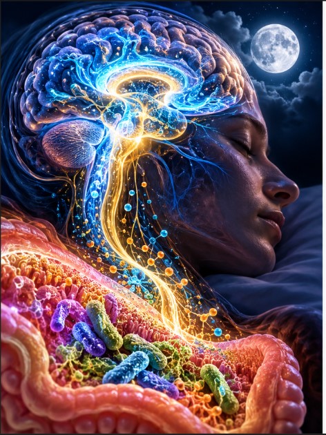
The gut is in constant conversation with the brain. Microbes rewrite the chemical environment in which we live. Nutrients become signals. Signals shape nerves. Nerves shape organs, immunity and sleep. And while we sleep, the brain enters a radically different physiological state—one that appears to power the movement of fluid and the clearance of certain metabolic by-products.
What happens when these systems stop working in harmony?
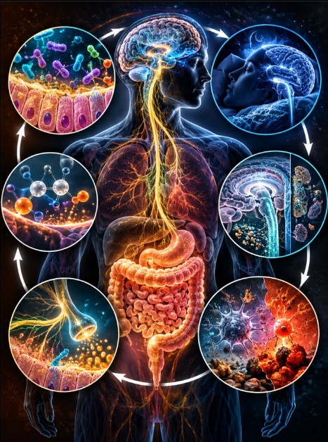
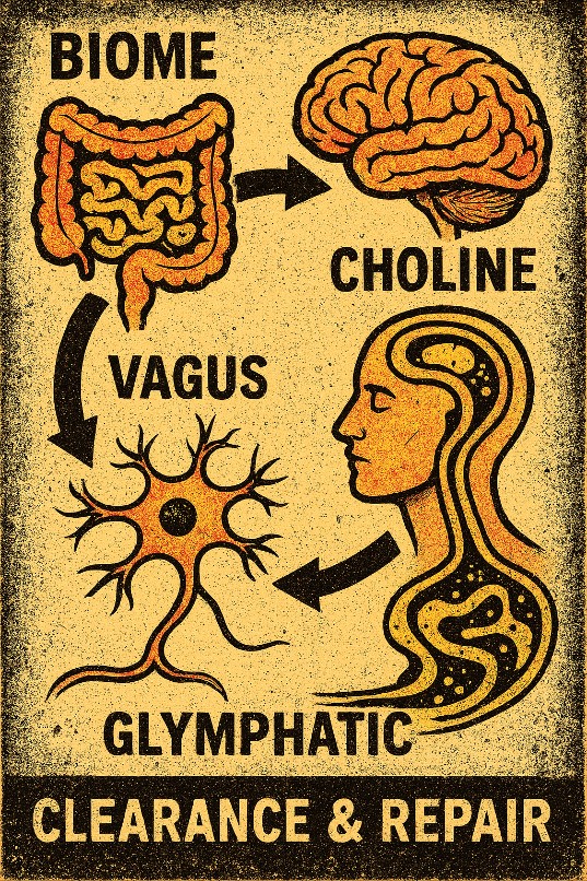
Our TCVG framework brings four pieces of this puzzle together: Terrain, Choline, Vagus and Glymphatic function. It proposes that a disturbance in one domain can cascade through the others, eventually locking the body into a biological state that is stubbornly difficult to reverse.
The most intriguing part is not any single component. It is the possibility that the components form a self-reinforcing feedback loop.
And in the year since this idea first took shape, the research world has delivered a cascade of findings that make the connections even more compelling—and the stakes higher.
1. It begins at the boundary
The colon is not simply a tube through which food passes. Its inner surface is a living, dynamic boundary between the outside world and the internal environment of the body.
A mucus layer helps separate much of the intestinal microbial community from the epithelial cells beneath it. Goblet cells produce mucus, epithelial cells regulate transport and barrier integrity, and microbial communities process the material that reaches them. Short-chain fatty acids, especially butyrate, fuel the energy supply and regulation of the intestinal lining.
Within this environment, three microbial groups have become particularly interesting to our work:
- Akkermansia, associated with mucin metabolism and interactions with the mucus barrier.
- Faecalibacterium, including F. prausnitzii, an important butyrate-producing commensal.
- Roseburia, another group of important fibre-fermenting, butyrate-producing bacteria.
We call them the Trio.
They are not the entire microbiome, nor do they perform identical jobs. Their importance lies in the combination of functions they represent: microbial interaction with mucus, fermentation of dietary substrates, production of metabolites, and participation in the ecological environment that supports intestinal homeostasis.
Think of the Trio less as three magic bacteria and more as three informative occupants of a much larger biological landscape—early-warning sensors of ecosystem health.
When that landscape changes, the consequences may extend well beyond the colon. Altered microbial activity can change the supply of metabolites, immune signals and chemical substrates available to the host. Barrier dysfunction can rewrite the relationship between microbes and the immune system. The host, in turn, changes the conditions under which the microbes live.
This is our first important principle: the terrain and the host are continually shaping one another.
It also means that microbial abundance alone tells only part of the story. What organisms are doing may matter just as much as which organisms are present. Recent multi-omics work in conditions ranging from schizophrenia to white-matter hyperintensities has begun linking reductions in butyrate-producing genera (including Faecalibacterium) to both cognitive measures and imaging proxies of brain fluid transport—exactly the kind of multi-system signal TCVG predicts.
2. Choline: a molecule with several jobs
Choline rarely receives the attention it deserves outside specialist research, yet it sits at the intersection of several fundamental biological processes.
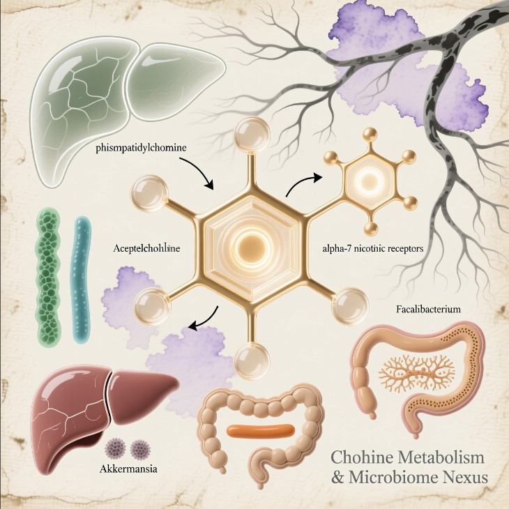
It contributes to phosphatidylcholine, an important component of cell membranes and bile. It is the precursor of acetylcholine, a neurotransmitter involved in neural and autonomic signalling. It can also be converted into betaine, which participates in one-carbon metabolism and methyl-group transfer.
In other words, choline is not simply a nutrient to tick off a dietary checklist. It is part of the chemical economy that supports membranes, metabolism and communication.
The host obtains choline from food and endogenous pathways, recycles it through normal physiology and distributes it among competing demands. The gut microbiome also changes the fate of dietary choline, including its conversion into microbial metabolites.
A 2025 review in Nature Metabolism brought together the expanding picture of choline metabolism across cellular and organism-wide functions, highlighting its roles in phospholipids, acetylcholine and betaine.
Then came an especially interesting development.
A 2026 study published in Nature reported that selected commensal bacteria can convert dietary choline into acetylcholine within the living gut environment. In experiments involving mice, colonisation with an acetylcholine-producing Bifidobacterium breve strain increased intestinal IgA production, altered microbial composition and improved resistance to an enteric infection. The researchers even identified the bacterial enzymes responsible and created a mutant strain that could no longer make the molecule—confirming the causal link.
That finding adds a fascinating dimension to the conversation: microbial metabolism can produce a molecule traditionally discussed as a host neurotransmitter, and that molecule can participate in local immune education.
It does not mean gut bacteria supply all the acetylcholine needed by the nervous system. It does show why the boundaries between microbial chemistry, host metabolism and biological signalling deserve a closer look.
Within TCVG, the question becomes whether changes in the microbial terrain can alter the availability or routing of choline-related metabolites in ways that influence other systems.
The relationship is not a simple equation in which fewer bacteria automatically mean less acetylcholine. Different pathways, tissues and microbial communities matter. But the metabolic bridge is real enough to investigate—and considerably more interesting than treating the gut and nervous system as unrelated subjects.
Trivia worth remembering: the same choline molecule that becomes membrane phospholipid in one cell can become a neurotransmitter in another, or a methyl donor in a third. The body is constantly negotiating these trade-offs.
3. The vagus: the body’s information highway
The vagus nerve is one of the major communication routes connecting internal organs with the brain.
It carries sensory information from the body towards the central nervous system and contributes to parasympathetic regulation of organs including the heart and gastrointestinal tract. It is part of a much larger network that includes other nerves, immune mediators, hormones and circulating metabolites.
This is not a one-way telephone line.
The brain can influence intestinal motility, secretion and immune activity. The gut can send information back through neural and non-neural routes. Microbial metabolites can interact with sensory pathways, while the host’s feeding patterns, stress responses and autonomic state help shape the intestinal environment.
A major 2025 review in Nature Reviews Microbiology described the gut–brain axis as a bidirectional communication network involving microbial signals, sensory epithelial cells and neural pathways.
The vagus also intersects with cholinergic regulation of inflammation. Acetylcholine can influence immune activity through specific receptor pathways, including the α7 nicotinic acetylcholine receptor in relevant experimental systems.
This creates a potentially important meeting point between the choline economy, autonomic regulation and immune control.
Our model asks what happens when these signals become persistently altered. Could changes in microbial function, metabolite availability and autonomic regulation reinforce one another? Could a system initially responding to a local disturbance settle into a different pattern of operation?
The significance is not that the vagus controls everything. It does not. The significance is that it offers one route through which local biological changes can become part of a wider physiological response.
Recent work has even shown that gut microbial ammonia can act as a compensatory signal that boosts colonic acetylcholine release and restores motility when host cholinergic signalling falters—another example of the host and microbiome negotiating in real time.
4. The night shift: the brain changes state
Sleep is not simply a period during which the brain switches off.
During sleep, neural activity, autonomic state, breathing, vascular dynamics and the movement of fluid through the brain all change. These coordinated shifts help create the conditions for processes involved in restoration and maintenance.
The glymphatic system is one of the most intriguing parts of this story.
It describes pathways through which cerebrospinal fluid (CSF) interacts with fluid in the brain’s tissue, including routes associated with blood vessels and astrocytes. Aquaporin-4, a water channel concentrated at astrocytic endfeet, is an important part of this architecture. Meningeal lymphatic vessels provide additional drainage routes connecting the central nervous system with peripheral lymphatic pathways.
The picture is more complex than a nightly rinse cycle. Fluid transport works alongside cellular uptake, degradation, protein regulation and immune surveillance. Different routes may handle different substances under different conditions.
But sleep appears to matter—a lot.
In January 2025, research published in Cell identified coordinated oscillations in norepinephrine, cerebral blood volume and CSF movement as strong predictors of glymphatic inflow and clearance during non-REM sleep in experimental systems. The researchers found that manipulating vascular dynamics could alter fluid transport, pointing to slow blood-vessel movements (vasomotion) as an active driver of the process. Optogenetic stimulation of the locus coeruleus—the brain’s norepinephrine factory—could even boost the pump.
This is an important shift in how we think about the brain’s night shift.
The story is not merely that sleep gives the brain time to clean up. The physiology of sleep appears to help generate the conditions that move fluid through the brain.
That makes sleep architecture, autonomic state and vascular rhythms part of the same conversation.
And it raises a much bigger question: what happens when those conditions are repeatedly disrupted? Certain common sleep medications have already been shown, in animal models, to suppress the very norepinephrine oscillations that drive the clearance pump—raising the possibility that some “solutions” for sleep may quietly undermine the night shift they are meant to support.
5. The human evidence is moving forward—fast
For years, much of the most detailed glymphatic research came from animal models. Human evidence has been harder to obtain, partly because fluid movement through the living brain is difficult to measure directly.
That picture is changing rapidly.
A 2026 study published in Nature Communications investigated sleep-associated glymphatic clearance of amyloid-beta and tau in humans. In a randomised crossover experiment involving 39 participants, the researchers compared normal sleep with sleep deprivation and observed higher morning plasma levels of Alzheimer’s-related biomarkers following normal sleep. Their interpretation, supported by additional physiological measurements and modelling, was consistent with sleep-active clearance contributing to the movement of these proteins out of the brain.
Why does this matter?
Because it moves the conversation beyond the assumption that glymphatic clearance is simply an interesting phenomenon observed in laboratory animals. It provides human evidence connecting sleep, physiological measurements and the handling of brain-associated proteins.
It does not settle every question about how much human glymphatic transport contributes to long-term disease. But it strengthens the case for investigating sleep-dependent clearance as a meaningful part of brain maintenance.
And then, in early 2026, a paper in the Journal of Affective Disorders examined relationships between gut microbiota and glymphatic-related imaging measures using bidirectional Mendelian-randomisation analyses. The researchers reported multiple causal associations and identified converging genetic signals involving the midbrain and glutamatergic neurons. Specific genera showed consistent negative associations with the DTI-ALPS index (a widely used proxy for perivascular water movement).
The study used the DTI-ALPS index as a proxy related to perivascular water movement; it was not a direct measurement of the entire glymphatic system. Its findings therefore need to be interpreted in the context of that measurement and the assumptions of the method.
Even so, the direction of the question is striking: gut microbial variation and measures related to brain fluid transport are now being examined within the same analysis—and causal arrows are beginning to appear.
Other 2025–2026 studies have linked glymphatic imaging markers to migraine, idiopathic intracranial hypertension, schizophrenia, and cognitive heterogeneity in white-matter disease, often alongside gut microbial signatures. The field is no longer waiting for perfect tools; it is using the best available proxies to ask systems-level questions.
6. The clock links the gut to the night
There is another piece that fits naturally into this architecture: circadian biology.
The gut has its own rhythms. Feeding, fasting, bile secretion, epithelial renewal, immune activity and microbial metabolism vary across the day. The central circadian clock helps coordinate these rhythms, while microbial metabolites can feed back into host signalling and peripheral clock regulation.
A 2025 review in Applied Microbiology and Biotechnology described this two-way relationship between circadian rhythms and the gut microbiome, including the influence of feeding patterns, microbial metabolites, immune signalling and peripheral clocks.
This matters because the brain’s maintenance processes are also state-dependent. Sleep timing, sleep quality, vascular dynamics and autonomic activity all influence the physiological conditions under which fluid transport occurs.
The gut and brain are therefore not merely connected by a nerve. They are embedded in a shared organism-wide timing system.
Imagine repeatedly disturbing feeding rhythms, microbial activity, sleep and autonomic balance. Each change may have a local effect. Together, they could alter the timing and coordination of several systems at once.
That is the kind of interaction a coupled model is designed to illuminate.
7. When maintenance becomes part of the problem
The brain produces metabolic by-products continuously. Its cells must regulate proteins, lipids, ions, energy use and inflammatory responses while maintaining the environment required for neural signalling.
Microglia, astrocytes, vascular cells and other components of the nervous system participate in this work. If inflammatory signals persist, oxidative stress rises or protein handling becomes abnormal, the consequences can extend beyond any single pathway.
Iron dysregulation and ferroptosis—the iron-dependent form of regulated cell death associated with lipid peroxidation—are being investigated in several neurological conditions. Protein aggregates, including amyloid-beta, tau and alpha-synuclein, are also central to different neurodegenerative processes.
These are not all interchangeable forms of biological waste, and they do not all arise from the same cause. Nor does their presence prove that glymphatic dysfunction initiated the problem.
But they illustrate why brain maintenance cannot be reduced to one molecule or one drainage route. The system must coordinate fluid movement, cellular metabolism, protein handling and immune regulation.
If several of those processes become disturbed together, the resulting environment may become increasingly difficult to stabilise.
This is where the idea of a persistent altered biological state becomes useful.
A disturbance may begin in one place and spread. The system may compensate. Those compensations may alter other pathways. Over time, the organism may operate in a different configuration from the one that existed before the original disturbance.
The question is no longer only, “What started the problem?”
It becomes, “What is now helping to keep it going?”
8. TCVG: the loop, not the domino line
The TCVG framework brings the story together through four interacting domains:
T — Terrain. The microbial ecosystem, mucus-associated organisms, fermentation, barrier function and immune environment.
C — Choline. The metabolic network connecting phosphatidylcholine, acetylcholine, betaine and microbial processing of dietary substrates.
V — Vagus. The neural and autonomic pathways that communicate information between the gut, brain and internal organs.
G — Glymphatic and brain maintenance. Sleep-dependent fluid transport, vascular dynamics, meningeal lymphatic drainage and the wider machinery that maintains the brain’s internal environment.
The proposed relationship is not a rigid sequence in which every person must begin with a depleted microbiome and end with a failed clearance system.
It is a feedback architecture.
A change in terrain may influence metabolites and host signalling. Those changes may interact with autonomic regulation. Autonomic state and sleep physiology may influence the conditions under which brain-maintenance processes operate. Meanwhile, altered sleep, stress physiology, feeding rhythms and gastrointestinal function can feed back into the gut environment.
The loop can potentially reinforce itself.
This is the central idea: the final expression of a biological disturbance may look very different from its starting point.
The first detectable change might be intestinal. The most consequential effects may emerge elsewhere. And by the time symptoms become visible, several systems may already be influencing one another.
TCVG offers a way to examine those relationships as one connected biological problem rather than a collection of unrelated observations.
9. Is the glymphatic collapse the final expression?
The title is deliberately provocative.
A glymphatic collapse would not mean that all fluid movement in the brain stops. It would mean that important aspects of the brain’s maintenance and clearance network become persistently impaired in a way that contributes to wider physiological dysfunction.
The evidence does not establish this as the universal endpoint of autism, ADHD, bipolar disorder, schizophrenia, Parkinson’s disease and Alzheimer’s disease. These conditions have distinct clinical identities, multiple contributing mechanisms and different trajectories.
But the broader question is compelling: could some conditions share disturbances in sleep, autonomic regulation, microbial signalling, metabolic function and brain maintenance, even when their causes and clinical presentations differ?
The emerging research makes that question increasingly interesting. Human sleep studies are advancing our understanding of brain-associated clearance. New work is examining microbial contributions to acetylcholine production. Circadian research is revealing more about the timing relationship between the gut and host physiology. Mendelian-randomisation and multi-omics studies are beginning to draw causal arrows between specific gut taxa and imaging proxies of brain fluid transport. And the molecular pump that drives the night shift—norepinephrine-mediated slow vasomotion—has now been identified and experimentally manipulated.
Individually, these findings do not establish the entire TCVG framework. Together, they open a much richer field of questions about how biological systems interact.
The brain does not maintain itself in isolation. It is part of an organism whose microbial ecology, metabolism, neural signalling, immune responses and daily rhythms are continually connected.
Perhaps the most important shift is to stop asking only which isolated component is broken and start asking what happens when the connections between components stop supporting recovery.
The glymphatic system may be one of the places where the consequences become visible.
But the story may begin much earlier—in the terrain, in the molecules that connect systems, and in the signals that determine whether the body can return to balance.
Different entry points. Interacting systems. A possible shared pattern of persistence.
That is the architecture we are investigating.
And the brain’s night shift may be one of its most revealing windows.
Selected research shaping this perspective
- Hauglund et al. (2025), Cell — norepinephrine-driven slow vasomotion and glymphatic clearance during sleep.
- Kenny et al. (2025), Nature Metabolism — cellular and organismal functions of choline metabolism.
- Ohara and Hsiao (2025), Nature Reviews Microbiology — microbiota–neuroepithelial signalling across the gut–brain axis.
- Nature Communications (2026) — human sleep-associated glymphatic clearance of amyloid-beta and tau.
- Journal of Affective Disorders (2026) — bidirectional Mendelian-randomisation analysis of gut microbiota and glymphatic-related imaging measures.
- Nature (2026) — commensal-derived acetylcholine and mucosal immune education.
- Bautista and colleagues (2025), Applied Microbiology and Biotechnology — bidirectional interactions between circadian rhythms and the gut microbiome.
The full evidence trail and mechanistic development behind the TCVG framework are available on request.