MICROGLIA: THE BRAIN’S IMMUNE CARETAKERS
When the Brain’s Maintenance System Changes Priorities
When microglia enter a sustained inflammatory or otherwise dysregulated state, their function can shift away from the balanced surveillance, maintenance and remodelling that normally support the neural environment. This does not mean that they simply stop working; rather, the signals governing their work may change, altering which functions are prioritised and how effectively they coordinate with surrounding cells. Because microglia participate in synaptic pruning, their altered signalling may affect the timing, selection or extent of neural connection refinement during development. Depending on the context, pruning may be excessive, insufficient or simply mistimed—and each could have different consequences for the developing network. The result is a compelling biological question: could altered microglial function, in some neurodevelopmental contexts, contribute to neural circuits being refined differently, with consequences for how information is processed, integrated and retrieved? This would not make microglia the cause of neurodivergence. It would make them one potential contributor to the biological environment in which neurodevelopment unfolds.
MICROGLIA: THE BRAIN’S IMMUNE CARETAKERS
How the cells that protect, maintain and shape the brain also help write the rules of learning, memory and communication — and why an inflamed, poorly cleared environment can change the script
The brain is not a pure neuron machine. It is a living tissue that must be continuously surveyed, cleaned, refined and defended. Microglia are the resident specialists for that job — the brain’s immune caretakers that spend their lives monitoring synapses, clearing debris and helping decide which neural connections stay and which go.
When their priorities shift — through inflammation, genetic risk factors, developmental timing, signals from the gut, or failures in the brain’s own waste-clearance system — the architecture of neural circuits can change. That does not make microglia the sole cause of neurodivergence. It does make them one of the most interesting biological players currently under the microscope, especially once we factor in two under-appreciated forces: persistent inflammatory messaging and the glymphatic clearance system discovered by Maiken Nedergaard’s group.
1. Meet the microglia: more than security guards
Neurons get the headlines. They fire, they wire, they compute. But they operate inside an environment shaped by glia. Microglia are the brain’s specialised immune cells, and they are anything but passive.
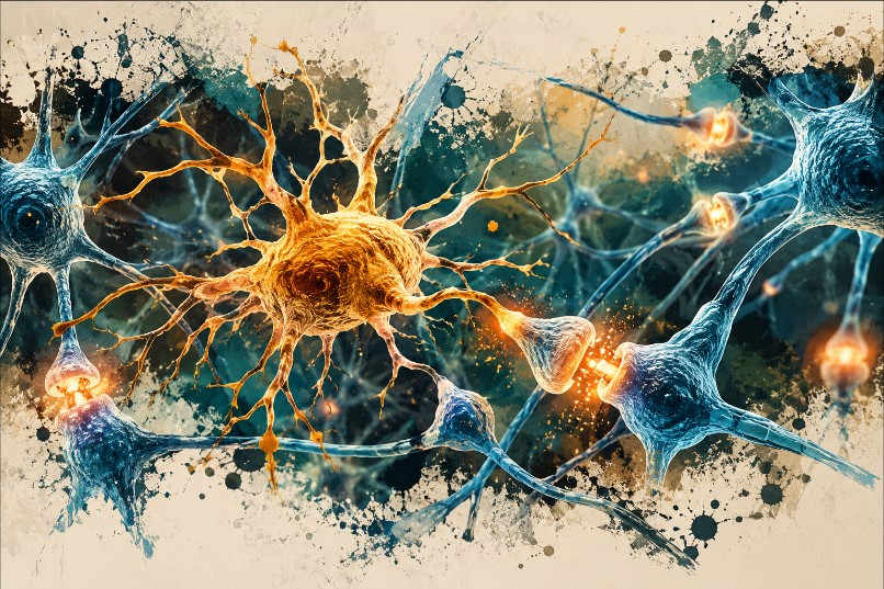
They constantly patrol tissue, sense subtle changes in chemistry and cellular health, remove material that needs clearing, and actively participate in synaptic remodelling across development and into adulthood. Their toolkit includes surveillance, debris clearance, synaptic pruning, immune signalling and local support for repair and adaptation.
They sit at the intersection of immunity and neural circuit construction. They do not think our thoughts, but they help maintain — and sometimes rewrite — the physical substrate in which those thoughts occur.
2. Why pruning matters: building a brain is not just about adding connections
A developing brain produces a vast excess of synapses. Refinement is essential. Some connections strengthen with use; others are weakened or eliminated. This is not random demolition. It is activity-dependent sculpting that helps networks specialise.
Microglia are key participants in this synaptic pruning. Think of a growing city that builds many roads and later optimises the network: useful routes are reinforced, redundant ones removed. Microglia help perform that optimisation, guided by molecular tags (including complement proteins) and local activity signals.
Recent work has sharpened the picture. In models carrying autism-associated SCN2A mutations, microglia show excessive phagocytic pruning of postsynaptic structures during specific developmental windows; removing or modulating those microglia can partially restore synaptic transmission and spine density. Parallel human cerebral organoid work with microglia incorporated shows the same over-pruning phenotype. A 2025 CRISPRi screen of autism-risk genes in human iPSC-derived microglia identified ADNP as a specific modifier of synaptic pruning, altering endocytic trafficking and microglia–neuron interactions.
These findings do not collapse every neurodevelopmental difference into “too much” or “too little” pruning. Timing, brain region, genetic background and co-occurring signals all matter. But they make clear that microglia are not bystanders in circuit construction — they are active editors.
3. When protection becomes part of the problem — the inflamed messaging cascade
Microglia respond to infection, injury, cellular stress and inflammatory signals. That responsiveness is protective. The difficulty arises when the response becomes sustained, poorly timed or mismatched to the actual threat.
This is where the inflamed messaging cascade becomes central. Persistent low-grade inflammation — whether driven by peripheral immune activation, microbial signals, sleep disruption or unresolved cellular stress — keeps microglia in a primed or reactive state. Cytokines, complement components, extracellular ATP and other danger signals form a self-reinforcing loop. Microglia release more inflammatory mediators, which further alter neighbouring neurons and astrocytes, which in turn generate more signals that keep microglia activated.
In this state, the normal balance of surveillance versus pruning can tip. Pruning may become excessive in some circuits, insufficient or mistimed in others. The result is not simply “inflammation equals damage.” It is a shift in the developmental and maintenance priorities of the brain’s caretaker cells.
4. The glymphatic system: when the brain’s cleaning crew falters
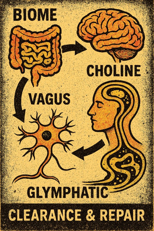
Enter the glymphatic system — the brain-wide fluid-transport network described by Nedergaard and colleagues. Cerebrospinal fluid flows along perivascular spaces, exchanges with interstitial fluid, and clears metabolic waste, proteins and inflammatory mediators, largely during sleep and driven in part by aquaporin-4 channels on astrocytic endfeet.
Glymphatic function and inflammation form a vicious cycle. Inflammatory signals and reactive gliosis can depolarise AQP4, disrupt perivascular flow and reduce clearance. Impaired clearance then allows cytokines, cellular debris and other signalling molecules to linger, further activating microglia and astrocytes. Microglia themselves appear to interact with this system; impaired meningeal lymphatic drainage can shift microglial phenotypes toward a more inflammatory profile.
Emerging imaging work already points to reduced glymphatic-related measures in autism, ADHD and other neurodevelopmental and psychiatric conditions, often co-occurring with the sleep disturbances that themselves impair clearance. The implication is straightforward and under-explored: a brain that cannot efficiently clear its inflammatory and metabolic load is a brain in which microglia are more likely to remain in a dysregulated state during the very windows when synaptic pruning is most active.
5. Biome depletion: the gut’s long reach into microglial maturation
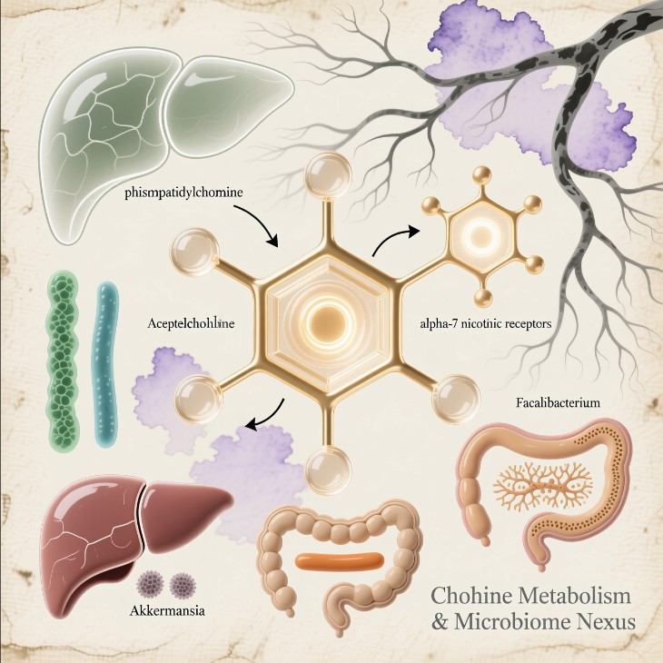
Microglia do not develop or operate in isolation from the rest of the body. Early-life gut microbiome composition is a powerful regulator of microglial maturation, density and functional state. Depletion or dysbiosis — whether from antibiotics, diet, maternal factors or other early insults — alters microglial gene expression, morphology and phagocytic behaviour.
Critical windows exist. Microbiota disruption in early life can produce enduring changes in microglial function into adolescence, including altered synaptic pruning and effects on learning-related plasticity. Short-chain fatty acids and other microbial metabolites influence microglial epigenetic state and inflammatory tone. When the microbiome is depleted or imbalanced, the signals that help keep microglia in a homeostatic, appropriately pruning mode are weakened.
This does not mean every case of neurodivergence is caused by a missing gut bacterium. It means that biome status is one of the upstream inputs that can calibrate the very cells responsible for refining neural circuits. Combine persistent inflammatory messaging with reduced glymphatic clearance and a depleted or dysbiotic microbiome, and the conditions for altered microglial pruning become more plausible.
6. ATP, purinergic signalling and the local alarm system
One of the most elegant signalling systems microglia use involves ATP. Inside cells, ATP is the universal energy currency. Outside cells, it becomes a potent messenger.
When ATP (or related purines) appears in the extracellular space — whether from stressed cells, inflammatory activity or impaired clearance — microglia detect it through P2X and P2Y receptors. That detection can trigger process motility, changes in inflammatory profile, and altered interaction with synapses. In an environment of persistent inflammation and slowed glymphatic clearance, extracellular signalling molecules are more likely to linger, amplifying the cascade.
7. How this constellation can influence learning, memory and neurodivergence
Learning and memory depend on the capacity of neural networks to change. Microglia shape that capacity through synaptic remodelling and the release of signalling molecules that affect plasticity. When microglia operate in a persistently inflamed, poorly cleared, microbially under-calibrated environment, the timing, selectivity and extent of pruning can shift.
In experimental models and emerging human cellular systems, this produces measurable differences in spine density, synaptic transmission and network function. The same logic extends to the heterogeneous biology of autism, ADHD and related developmental differences: not a single microglial lesion, but a set of conditions under which microglial editing of circuits becomes less precise.
8. Suramin and the purinergic hypothesis — still an open door
The 2017 small pilot trial of low-dose suramin and the subsequent larger study (which missed its primary endpoint but showed exploratory signals) keep the purinergic pathway on the table. An ongoing Phase 2 trial continues to examine it. Suramin is a blunt tool and is not an approved treatment. What it highlights is that modulating the extracellular signalling environment that microglia read remains a legitimate research question — especially once we recognise how inflammation, clearance failure and microbial signals all converge on that same environment.
9. What this integrated view suggests
Microglia are essential for normal brain maintenance, defence and developmental refinement. When their activity is appropriately timed and coordinated, they support healthy circuit construction. When that coordination is disrupted by a persistent inflammatory messaging cascade, impaired glymphatic clearance, and early biome depletion or dysbiosis, the resulting differences in synaptic architecture become a biologically plausible contributor to neurodevelopmental variation.
For parents and clinicians the practical foundation remains unchanged: developmental assessment, individualised educational and therapeutic support, and attention to sleep, hearing, seizures and other medical issues. At the same time, the science is converging on a richer picture. Sleep quality matters because it drives glymphatic clearance. Early microbial exposures matter because they calibrate microglia. Inflammatory tone matters because it can lock microglia into a state that changes how they prune.
The goal is not to silence the brain’s immune system or to declare any single pathway “the cause.” It is to understand how the protective, maintenance and sculpting functions of microglia are coordinated with the brain’s clearance systems and the body’s microbial signals — and what becomes possible when we learn to keep that coordination on track during the windows that matter most.
Selected references
Core pruning and microglia literature (SCN2A, ADNP CRISPRi, Mordelt & de Witte 2023, etc.).
Nedergaard glymphatic work and subsequent reviews on glymphatic–inflammation interactions.
Gut–microglia–pruning studies linking early dysbiosis to altered microglial maturation and synaptic refinement.
Emerging glymphatic imaging findings in neurodevelopmental conditions.
Suramin pilot and follow-up trials, plus ongoing Phase 2 work.
This article is an exploratory synthesis for educational purposes. It does not constitute medical advice, diagnosis or treatment recommendation.